
- Panel Features
- Panels List
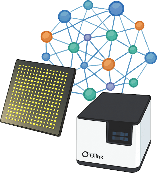
- Workflow
- Demo
- Case
- Why Creative Proteomics
- Sample Requirements
- Sample Submission Pack
What is the Olink Target 96 Immuno-Oncology Panel
The Olink Target 96 Immuno-Oncology Panel is a high-performance protein analysis tool designed to assess 92 immune-related biomarkers in cancer research. It requires only 1μL of biological sample, such as plasma or serum, and delivers results in relative units (NPX).
Features of the panel
- Comprehensive biomarker coverage: This panel analyzes 92 immune-related proteins, addressing key processes in cancer immunology such as tumor immunity, chemotaxis, vascular and tissue remodeling, apoptosis, cell killing, metabolism, and autophagy.
- Precise and reliable technology: The panel utilizes Proximity Extension Assay (PEA) and quantitative PCR to provide accurate and reproducible results with proven specificity and sensitivity across a wide dynamic range.
- Minimal sample requirement: Only 1µL of biological sample (plasma, serum, etc.) is required for analysis, enabling high-throughput studies with limited sample volume.
- Wide application potential: Ideal for research in immuno-oncology and related fields, it supports in-depth analysis of immune responses, tumor-associated processes, and cancer mechanisms.
List of 92 Immuno-Oncology Proteins
Protein category
The Olink Target 96 Immuno-Oncology Panel covers a broad range of proteins, including cytokines, chemokines, growth factors, enzymes, adhesion molecules, signaling molecules, and immune checkpoint proteins. These proteins are involved in various biological processes such as immune regulation, inflammation, cell migration, tumor biology, and immune cell signaling.
Table. List of Olink Target 96 Immuno-Oncology Panel
| Protein Category | UniProt ID | Gene | Protein Name |
| C-C Motif Chemokines | Q99616 | CCL13 | C-C motif chemokine 13 |
| P10147 | CCL3 | C-C motif chemokine 3 | |
| Q99731 | CCL19 | C-C motif chemokine 19 | |
| P80075 | CCL8 | C-C motif chemokine 8 | |
| P13236 | CCL4 | C-C motif chemokine 4 | |
| P80098 | CCL7 | C-C motif chemokine 7 | |
| P13500 | CCL2 | C-C motif chemokine 2 | |
| P55773 | CCL23 | C-C motif chemokine 23 | |
| Q92583 | CCL17 | C-C motif chemokine 17 | |
| P78556 | CCL20 | C-C motif chemokine 20 | |
| C-X-C Motif Chemokines | O14625 | CXCL11 | C-X-C motif chemokine 11 |
| Q07325 | CXCL9 | C-X-C motif chemokine 9 | |
| P10145 | CXCL8 | Interleukin-8 | |
| P09341 | CXCL1 | Growth-regulated alpha protein | |
| P02778 | CXCL10 | C-X-C motif chemokine 10 | |
| P42830 | CXCL5 | C-X-C motif chemokine 5 | |
| O43927 | CXCL13 | C-X-C motif chemokine 13 | |
| P48061 | CXCL12 | Stromal cell-derived factor 1 | |
| TNF Superfamily | P50591 | TNFSF10 | Tumor necrosis factor ligand superfamily member 10 |
| O75509 | TNFRSF21 | Tumor necrosis factor receptor superfamily member 21 | |
| O43557 | TNFSF14 | Tumor necrosis factor ligand superfamily member 14 | |
| O43508 | TNFSF12 | Tumor necrosis factor ligand superfamily member 12 | |
| Q07011 | TNFRSF9 | Tumor necrosis factor receptor superfamily member 9 | |
| P43489 | TNFRSF4 | Tumor necrosis factor receptor superfamily member 4 | |
| P01375 | TNF | Tumor necrosis factor | |
| Interleukins (IL) | P60568 | IL2 | Interleukin-2 |
| Q14116 | IL18 | Interleukin-18 | |
| P13232 | IL7 | Interleukin-7 | |
| P40933 | IL15 | Interleukin-15 | |
| P05231 | IL6 | Interleukin-6 | |
| P22301 | IL10 | Interleukin-10 | |
| P05113 | IL5 | Interleukin-5 | |
| P01583 | IL1A | Interleukin-1 alpha | |
| O95760 | IL33 | Interleukin-33 | |
| P35225 | IL13 | Interleukin-13 | |
| P29459_P29460 | IL12A_IL12B | Interleukin-12 | |
| P05112 | IL4 | Interleukin-4 | |
| Growth Factors | P09038 | FGF2 | Fibroblast growth factor 2 |
| P01127 | PDGFB | Platelet-derived growth factor subunit B | |
| P15692 | VEGFA | Vascular endothelial growth factor A | |
| P49763 | PGF | Placenta growth factor | |
| P14210 | HGF | Hepatocyte growth factor | |
| Q15389 | ANGPT1 | Angiopoietin-1 | |
| O15123 | ANGPT2 | Angiopoietin-2 | |
| P01133 | EGF | Pro-epidermal growth factor | |
| P09237 | MMP7 | Matrilysin | |
| P39900 | MMP12 | Macrophage metalloelastase | |
| Receptors and Surface Proteins | P06127 | CD5 | T-cell surface glycoprotein CD5 |
| P01730 | CD4 | T-cell surface glycoprotein CD4 | |
| P01732 | CD8A | T-cell surface glycoprotein CD8 alpha chain | |
| P10747 | CD28 | T-cell-specific surface glycoprotein CD28 | |
| P25942 | CD40 | Tumor necrosis factor receptor superfamily member 5 | |
| P29965 | CD40LG | CD40 ligand | |
| P26842 | CD27 | CD27 antigen | |
| P32970 | CD70 | CD70 antigen | |
| Q9NZQ7 | CD274 | Programmed cell death 1 ligand 1 | |
| Q01151 | CD83 | CD83 antigen | |
| Q9BZW8 | CD244 | Natural killer cell receptor 2B4 | |
| Q13241 | KLRD1 | Natural killer cells antigen CD94 | |
| P42701 | IL12RB1 | Interleukin-12 receptor subunit beta-1 | |
| Q02763 | TEK | Angiopoietin-1 receptor | |
| Granzyme and Cytotoxic Proteins | P20718 | GZMH | Granzyme H |
| P12544 | GZMA | Granzyme A | |
| P10144 | GZMB | Granzyme B | |
| P48023 | FASLG | Tumor necrosis factor ligand superfamily member 6 | |
| Other Immunomodulatory and Signaling Proteins | P00813 | ADA | Adenosine deaminase |
| P29474 | NOS3 | Nitric oxide synthase | |
| P35968 | KDR | Vascular endothelial growth factor receptor 2 | |
| P18627 | LAG3 | Lymphocyte activation gene 3 protein | |
| P09601 | HMOX1 | Heme oxygenase 1 | |
| Q29983_Q29980 | MICA_MICB | MHC class I polypeptide-related sequence A and MHC class I polypeptide-related sequence B | |
| Q9Y653 | ADGRG1 | Adhesion G-protein coupled receptor G1 | |
| P09603 | CSF1 | Macrophage colony-stimulating factor 1 | |
| O95727 | CRTAM | Cytotoxic and regulatory T-cell molecule | |
| P01579 | IFNG | Interferon gamma | |
| Miscellaneous Proteins | P07585 | DCN | Decorin |
| O00182 | LGALS9 | Galectin-9 | |
| P09382 | LGALS1 | Galectin-1 | |
| O76036 | NCR1 | Natural cytotoxicity triggering receptor 1 | |
| P43629 | KIR3DL1 | Killer cell immunoglobulin-like receptor 3DL1 | |
| Q16790 | CA9 | Carbonic anhydrase 9 | |
| P21246 | PTN | Pleiotrophin | |
| Q9UQV4 | LAMP3 | Lysosome-associated membrane glycoprotein 3 | |
| Q14790 | CASP8 | Caspase-8 | |
| P78423 | CX3CL1 | Fractalkine | |
| O75144 | ICOSLG | ICOS ligand | |
| Other Proteins | Q8WXI7 | MUC16 | Mucin-16 |
| Q15116 | PDCD1 | Programmed cell death protein 1 | |
| P32970 | CD70 | CD70 antigen | |
| P05089 | ARG1 | Arginase-1 | |
| P01137 | TGFB1 | Transforming growth factor beta-1 proprotein | |
| Q9NP84 | TNFRSF12A | Tumor necrosis factor receptor superfamily member 12A | |
| Q9BQ51 | PDCD1LG2 | Programmed cell death 1 ligand 2 |
Protein Functions
Biological process
The 92 proteins are associated with biological processes such as apoptosis, autophagy, metabolism/autophagy, promotion of tumor immunity, inhibition of tumor immunity, as well as vascular and tissue remodeling.

Diseases
The 92 proteins are widely applied in the fields of cancer, cardiovascular diseases, inflammation, infections, vascular and lymphatic systems, digestion, bone, and kidney diseases.

KEGG Pathways
An analysis of the most significantly enriched pathways (p < 0.05) revealed that the pathway with the highest enrichment of the 92 proteins was Cytokine-cytokine receptor interaction, followed by Rheumatoid arthritis and Chemokine signaling pathway.

Workflow of Olink Target 96 Immuno-Oncology Panel

Demo Results of Olink Data
(Figures come from Liu, J., et al. 2022)

Heatmap of plasma protein of all samples by olink target 96 immuno-oncology panel.

Statistical comparison of IL-18 NPX measurement values.
Case Study

A Targeted Proteomics Approach Reveals a Serum Protein Signature as a Diagnostic Biomarker for Colorectal Cancer
Journal: Journal of inflammation research
Year: 2024
- Background
- Results
Colorectal cancer (CRC) is showing an increasing incidence and mortality rate in China, with approximately 555,000 new cases and 286,000 deaths reported in 2020. Early symptoms of CRC are often not apparent, and most patients are diagnosed at advanced stages. Early detection and targeted therapy are crucial for improving survival rates. The study aims to identify non-invasive biomarkers for CRC by detecting changes in tumor immune-related circulating proteins in peripheral blood.
Ninety-two proteins were detected in the serum of CRC patients' peripheral blood, and 49 differentially expressed proteins were identified, of which 42 were upregulated and 7 were downregulated. GO analysis revealed that genes with high expression levels in CRC were mainly enriched in cell adhesion and lymphocyte proliferation signaling pathways. KEGG enrichment analysis showed that these genes were primarily enriched in cytokine-cytokine receptor interactions and the PI3K-AKT signaling pathway.
 Figure 1. Differential protein expression heat map. (Wan, Y., et al., 2024)
The differential proteins identified were used to predict a signature model, which revealed that a signature composed of 6 proteins (IL7, CXCL12, IL10, IL15, CXCL1, and MCP-3) could effectively differentiate CRC from control samples. The performance of the signature model was evaluated using Receiver Operating Characteristic (ROC) analysis, showing an AUC of 0.9224 in the training set and 0.8992 in the total set. The performance surpassed that of the classic clinical markers CEA and CA-19-9.
 Figure 2. Prediction result of protein signature model. (Balzer, M. S., et al., 2024)
Subsequently, the best protein model identified was validated in CRC and control samples using ELISA technology, with IL7, CXCL12, IL10, and CXCL1 showing consistent results with the Olink findings. Impedance analysis was used to measure the absolute counts of immune cells (lymphocytes, monocytes, neutrophils, eosinophils, and basophils) in the peripheral blood of CRC patients. The results showed that the absolute count of lymphocytes in the CRC group was lower than that in the control group, while the absolute count of monocytes in CRC patients was significantly higher than in healthy controls. CXCL1 levels were positively correlated with the absolute count of basophils, and IL10 levels were positively correlated with the absolute count of neutrophils.
Why Creative Proteomics
Comprehensive After-Sales Support
We offer complete support from experimental design to data analysis, ensuring smooth progress.
Expert Team and Technical Support
Our experienced team provides personalized technical support to optimize your research.
High-Quality Experimentation and Data Management
We ensure high-quality sample handling and data analysis for reliable results.
Customized Post-Analysis and Reporting
We provide tailored reports and expert guidance to help you interpret and apply your findings effectively.
Sample Requirements
| Sample Type | Recommended Sample Size | Sample Quality | Pre-treatment and Storage | Sample Transport |
| Plasma/Serum/Body Fluid | 40µL/sample | Protein concentration: 0.5mg/ml ~ 1mg/ml | Transfer to a clean tube, aliquot into EP tubes or 96-well plates, store at -80℃ | Seal with foil, ship with dry ice |
| Tissue | ||||
| Cells | ||||
| Exosomes | ||||
| Other |
References
- Liu, J., Wang, Y., Tian, Z., et al. (2022). Multicenter phase II trial of Camrelizumab combined with Apatinib and Eribulin in heavily pretreated patients with advanced triple-negative breast cancer. Nature communications, 13(1), 3011. https://doi.org/10.1038/s41467-022-30569-0
- Wan, Y., Luo, W., Song, X., Zhao, Y., Han, Z., Shen, J., Xie, F., Li, Y., & He, J. (2024). A Targeted Proteomics Approach Reveals a Serum Protein Signature as a Diagnostic Biomarker for Colorectal Cancer. Journal of inflammation research, 17, 10755–10768. https://doi.org/10.2147/JIR.S492356