
- Panel Features
- Panels List
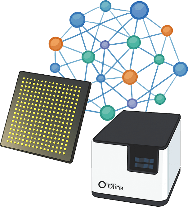
- Workflow
- Demo
- Case
- FAQ
- Why Creative Proteomics
- Sample Requirements
- Sample Submission Pack
What is the Olink Target 96 Oncology III Panel
Customized panel for human
The Olink Target 96 Oncology III Panel quantifies proteins, with detailed biomarker information available on the Olink website. Utilizing Olink® Biomarker technology (Olink®, Uppsala, Sweden), 92 proteins were analyzed. The process involves three stages: incubation, extension/amplification, and detection. During incubation, DNA-labeled antibody pairs are introduced to the sample and incubated overnight to bind target proteins. The following day, extension and amplification generate unique DNA reporter sequences for each protein, followed by preamplification via standard PCR. Detection employs high-throughput real-time qPCR on the Olink Signature Q100 System to measure DNA reporter sequences. Samples were randomly distributed across plates. Data underwent quality control and normalization using internal extension and interplate controls, accounting for intra- and inter-batch variability. Protein levels are reported as normalized protein expression (NPX) values, calculated on a log2 scale.
Features of the pane
- Species: primarily validated for human proteins, with no guaranteed cross-reactivity across other species
- Proteins: the simultaneous analysis of 92 protein biomarkers
- Sample: Using just 1µL of plasma, serum, or other compatible samples.
- Readout: Data are delivered in normalized protein expression (NPX) units, offering precise insights into relative protein abundance.
- Platform: The panel is designed to run on the Olink Signature Q100 system.
List of 92 human derived biomarkers
Protein category
The Olink Target 96 Oncology III Panel includes 92 proteins categorized into seven main groups: the enzymes (18), Receptors (10), Structural/Adhesion (12), Signaling Proteins (15), Immune-Related Proteins (10), Transport/Secretion (10), and other functional proteins (17). Each protein has been carefully selected by experts in the field and is involved in angiogenesis, cell communication, cell metabolism, apoptosis, cell proliferation/differentiation, and includes multiple markers of carcinogenicity, tumour progression, solid tumours, and recurrent tumours according to DisGenNet's inclusion of cancer-related proteins. A number of identified cancer-related biomarkers have also been added to the tumour III panel, including: cell surface A33 antigen, B-cell receptor CD22, proneuropeptide γ, allogeneic graft inflammatory factor 1, and vascular endothelial growth factor receptor 1.
Table. List of Olink Target 96 Oncology II I Panel
| Protein Category | UniProt ID | Gene | Protein Name |
| Enzymes | Q9H3G5 | CPVL | Probable serine carboxypeptidase CPVL |
| Q9H0P0 | NT5C3A | Cytosolic 5'-nucleotidase 3A | |
| P15121 | AKR1B1 | Aldo-keto reductase family 1 member B1 | |
| P28838 | LAP3 | Cytosol aminopeptidase | |
| P49441 | INPP1 | Inositol polyphosphate 1-phosphatase | |
| P51580 | TPMT | Thiopurine S-methyltransferase | |
| P55789 | GFER | FAD-linked sulfhydryl oxidase ALR | |
| P06865 | HEXA | Beta-hexosaminidase subunit alpha | |
| P08397 | HMBS | Porphobilinogen deaminase | |
| P09110 | ACAA1 | 3-ketoacyl-CoA thiolase | |
| Q96I15 | SCLY | Selenocysteine lyase | |
| Q14353 | GAMT | Guanidinoacetate N-methyltransferase | |
| Q9Y5K2 | KLK4 | Kallikrein-4 | |
| Q9H773 | DCTPP1 | dCTP pyrophosphatase 1 | |
| P43490 | NAMPT | Nicotinamide phosphoribosyltransferase | |
| P48643 | CCT5 | T-complex protein 1 subunit epsilon | |
| P02771 | AFP | Alpha-fetoprotein | |
| P05187 | ALPP | Alkaline phosphatase | |
| Receptors | P17948 | FLT1 | Vascular endothelial growth factor receptor 1 |
| P20273 | CD22 | B-cell receptor CD22 | |
| P38484 | IFNGR2 | Interferon gamma receptor 2 | |
| P36888 | FLT3 | Receptor-type tyrosine-protein kinase FLT3 | |
| P13747 | HLA-E | HLA class I histocompatibility antigen | |
| P02745 | C1QA | Complement C1q subcomponent subunit A | |
| Q496F6 | CD300E | CMRF35-like molecule 2 | |
| Q14773 | ICAM4 | Intercellular adhesion molecule 4 | |
| Q9UMF0 | ICAM5 | Intercellular adhesion molecule 5 | |
| Q9NTU7 | CBLN4 | Cerebellin-4 | |
| Structural/Adhesion | Q9BYE9 | CDHR2 | Cadherin-related family member 2 |
| P20849 | COL9A1 | Collagen alpha-1(IX) chain | |
| P32004 | L1CAM | Neural cell adhesion molecule L1 | |
| P33241 | LSP1 | Lymphocyte-specific protein 1 | |
| Q92832 | NELL1 | Protein kinase C-binding protein NELL1 | |
| Q6PCB0 | VWA1 | von Willebrand factor A domain-containing protein 1 | |
| Q08174 | PCDH1 | Protocadherin-1 | |
| Q99795 | GPA33 | Cell surface A33 antigen | |
| Q9Y2B0 | CNPY2 | Protein canopy homolog 2 | |
| Q9UDT6 | CLIP2 | CAP-Gly domain-containing linker protein 2 | |
| Q9P1Z2 | CALCOCO1 | Calcium-binding and coiled-coil domain-containing protein 1 | |
| Q99536 | VAT1 | Synaptic vesicle membrane protein VAT-1 homolog | |
| Signaling Proteins | P20340 | RAB6A | Ras-related protein Rab-6A |
| P30260 | CDC27 | Cell division cycle protein 27 homolog | |
| P42331 | ARHGAP25 | Rho GTPase-activating protein 25 | |
| P42575 | CASP2 | Caspase-2 | |
| P62166 | NCS1 | Neuronal calcium sensor 1 | |
| O43524 | FOXO3 | Forkhead box protein O3 | |
| O43707 | ACTN4 | Alpha-actinin-4 | |
| O60542 | PSPN | Persephin | |
| O60907 | TBL1X | F-box-like/WD repeat-containing protein TBL1X | |
| Q93096 | PTP4A1 | Protein tyrosine phosphatase type IVA 1 | |
| P28827 | PTPRM | Receptor-type tyrosine-protein phosphatase mu | |
| Q13459 | MYO9B | Unconventional myosin-IXb | |
| Q9Y5K8 | ATP6V1D | V-type proton ATPase subunit D | |
| Q9UJY5 | GGA1 | ADP-ribosylation factor-binding protein GGA1 | |
| Q9H8J5 | MANSC1 | MANSC domain-containing protein 1 | |
| Immune-Related Proteins | P01584 | IL1B | Interleukin-1 beta |
| P01591 | JCHAIN | Immunoglobulin J chain | |
| P13747 | HLA-E | HLA class I histocompatibility antigen | |
| P02745 | C1QA | Complement C1q subcomponent subunit A | |
| Q96PD4 | IL17F | Interleukin-17F | |
| Q14773 | ICAM4 | Intercellular adhesion molecule 4 | |
| Q9UMF0 | ICAM5 | Intercellular adhesion molecule 5 | |
| Q9UDT6 | CLIP2 | CAP-Gly domain-containing linker protein 2 | |
| Q9NTU7 | CBLN4 | Cerebellin-4 | |
| Q9H8J5 | MANSC1 | MANSC domain-containing protein 1 | |
| Transport/Secretion | Q9HD26 | GOPC | Golgi-associated PDZ and coiled-coil motif-containing protein |
| O60763 | USO1 | General vesicular transport factor p115 | |
| Q8NEZ2 | VPS37A | Vacuolar protein sorting-associated protein 37A | |
| Q5VIR6 | VPS53 | Vacuolar protein sorting-associated protein 53 homolog | |
| Q7Z739 | YTHDF3 | YTH domain-containing family protein 3 | |
| Q7Z5L0 | VMO1 | Vitelline membrane outer layer protein 1 homolog | |
| Q9Y5K8 | ATP6V1D | V-type proton ATPase subunit D | |
| Q9UJY5 | GGA1 | ADP-ribosylation factor-binding protein GGA1 | |
| Q9UDT6 | CLIP2 | CAP-Gly domain-containing linker protein 2 | |
| Q9H8J5 | MANSC1 | MANSC domain-containing protein 1 | |
| Miscellaneous | Q9BSL1 | UBAC1 | Ubiquitin-associated domain-containing protein 1 |
| Q9BS26 | ERP44 | Endoplasmic reticulum resident protein 44 | |
| Q9Y644 | RFNG | Beta-1 | |
| Q12904 | AIMP1 | Aminoacyl tRNA synthase complex-interacting multifunctional protein 1 | |
| P35637 | FUS | RNA-binding protein FUS | |
| P13693 | TPT1 | Translationally-controlled tumor protein | |
| P12872 | MLN | Promotilin | |
| O14558 | HSPB6 | Heat shock protein beta-6 | |
| O60575 | SPINK4 | Serine protease inhibitor Kazal-type 4 | |
| O75695 | RP2 | Protein XRP2 | |
| O76038 | SCGN | Secretagogin | |
| O95379 | TNFAIP8 | Tumor necrosis factor alpha-induced protein 8 | |
| P13521 | SCG2 | Secretogranin-2 | |
| O95715 | CXCL14 | C-X-C motif chemokine 14 | |
| P0DN86 | CGB3 | Choriogonadotropin subunit beta 3 | |
| P01303 | NPY | Pro-neuropeptide Y | |
| Q96FQ6 | S100A16 | Protein S100-A16 |
Protein Functions
Biological process
Primarily associated with metsbolic, carsiovascula, cancer, imuune, and neurological.

Disease area
Primarily associated with immune systerm diseases, signal transduction, metabolism, and innate immune system.

Workflow of Olink Proteomics

Demo Results of Olink Data
(Figures come from Ding, R., et al. 2024)

The bar chart displayed the number of proteins.

Volcano plots of differentially expressed proteins.

Heatmap of differentially expressed proteins.
Case Study

Proteomic and serological markers for diagnosing cardia gastric cancer and precursor lesions in a Chinese population
Journal: scientific reports
Year: 2024
- Background
- Results
Cardia cancer (CGC) accounted for 18% of global gastric cancer cases in 2018. Endoscopy remains the mainstay of diagnosis in high-incidence areas, but its invasiveness and high cost limit its wider application. This highlights the urgent need for non-invasive and cost-effective alternatives to enhance early detection and improve intervention outcomes in at-risk populations. Recent studies have demonstrated the effectiveness of serological markers such as pepsinogen (PG), gastrin-17 (G-17), and Helicobacter pylori (h.pylori) antibodies in risk stratification of non-cardia gastric cancer (NCGC) in high-prevalence countries such as China, Japan, and South Korea. These markers are significantly associated with atrophic gastritis and have been included in endoscopic screening protocols. However, their role in CGC remains to be further studied.
Analysis of expression profiles across 92 proteins in Healthy, CLGD, CHGD, and CGC groups demonstrated progressive changes in protein expression patterns with disease progression, as visualized in the cluster heatmap (Fig. 1A). Both dimensionality reduction techniques - t-SNE (Fig. 1B) and UMAP (Fig. 1C) - consistently revealed distinct clustering patterns, with the Healthy group forming a separate cluster in low-dimensional space, clearly distinguishable from the CLGD, CHGD, and CGC groups. The above results show that H. pylori type I and type II H. pylori positivity was significantly associated with CGC and precancerous lesions compared with H. pylori negative.
 Figure 1. Protein profiles differentiate CGC and precancerous states. (Jiqing Li, et al. 2024 )
FAQs
Do you have panels for other species, such as mice?
Our company offers two products for mouse proteins. The Target 96 Mouse Exploratory and Target 48 Mouse Cytokine Combinations have shown that the Target 96 Mouse Exploratory Panel is also suitable for the analysis of rat proteins. For more information, please contact technical support and refer to the technical note in the documentation centre, "Extending Olink's Mouse Exploratory Panel to Rat Studies". In addition to the mouse panel mentioned above, all Olink assays were developed using antibodies against human proteins. There is, of course, some degree of antibody cross-reactivity between human proteins and homologs of other species. As a result, a percentage of the assays in our panel produce signals in samples of non-human origin, and some of our customers are already using them in animal studies. Olink's human panel is only for human samples, but you can certainly use it for samples of non-human species at your own discretion.
Does Olink offer rodent-specific panels?
Olink offers a product for mouse proteins. The Target 96 Mouse Exploratory have shown that the Target 96 Mouse Exploratory Panel is also suitable for the analysis of rat proteins. For more information, please refer to our technical note "Extending Olink's Mouse Exploratory Panel to Rat Studies" or contact technical support.
Why Creative Proteomics
Advanced Bioinformatics Solutions
We provides advanced bioinformatics for Olink data analysis, delivering actionable research insights.
Diverse Scientific Applications
We supports disease modeling, biomarker discovery, and translational research across diverse scientific fields.
Efficient Workflow with High Precision
We uses Olink Q100 for precise, efficient sample analysis, ensuring reliable, reproducible research outcomes.
Comprehensive Research Support
We delivers end-to-end assistance, from experimental design to data analysis, ensuring smooth research processes with expert advice and resources to enable transformative discoveries.
Sample Requirements
| Sample Type | Recommended Sample Size | Sample Quality | Pre-treatment and Storage | Sample Transport |
| Plasma/Serum/Body Fluid | 40µL/sample | Protein concentration: 0.5mg/ml ~ 1mg/ml | Transfer to a clean tube, aliquot into EP tubes or 96-well plates, store at -80℃ | Seal with foil, ship with dry ice |
| Tissue | ||||
| Cells | ||||
| Exosomes | ||||
| Other |
References
- Li, J., Zhao, W., Yang, J.,et al. (2024). Proteomic and serological markers for diagnosing cardia gastric cancer and precursor lesions in a Chinese population. Scientific reports, 14(1), 25309. https://doi.org/10.1038/s41598-024-75912-1
- Fontanilles, M., Heisbourg, J. D., Daban, et al. (2024). Metabolic remodeling in glioblastoma: a longitudinal multi-omics study. Acta neuropathologica communications, 12(1), 162. https://doi.org/10.1186/s40478-024-01861-5
- Ding, R., Wu, L., Wei, S.,et.al. (2024). Multi-targeted olink proteomics analyses of cerebrospinal fluid from patients with aneurysmal subarachnoid hemorrhage. Proteome science, 22(1), 11. https://doi.org/10.1186/s12953-024-00236-x