
A common pre-study question sounds simple: "I have a candidate biomarker—can Olink detect the isoform I care about?" In practice, Olink isoform detection is rarely a yes/no. It depends on what the assay's binding reagents recognize: a protein target, a protein region, or a sequence feature that may be shared across multiple isoforms.
For most biomarker studies, that nuance is manageable—if you interpret results as an assay-defined protein signal and avoid isoform-specific claims unless you can support them.
This article explains:
- what an Olink assay reports (and what it does not)
- how Olink NPX interpretation stays manuscript-safe
- when isoforms and protein target specificity become limiting
- what to check before making isoform-level statements
- when Olink biomarker validation should include orthogonal methods (targeted MS, western blot, ELISA)
- what to ask before finalizing a panel
Introduction: Why Protein Target Interpretation Matters in Olink Studies
Start with the real technical question
If you already have a protein target (or a short candidate list), you are typically trying to answer one of two questions:
- Does the assay signal map cleanly to the molecular form I care about? (isoform/proteoform question)
- If it doesn't, can I still use the signal to prioritize biology and follow up correctly? (interpretation plan)
The practical issue is that the label in a panel list ("Protein X") is not the same as an experimentally proven statement about isoforms.
Why the answer is not always a simple yes or no
Isoform interpretation depends on variables you can actually check:
- the assay's target region / binding site (if available)
- isoform structure (shared vs unique regions)
- specificity evidence and likely cross-reactivity
- whether isoform distinction is biologically necessary
What Does an Olink Assay Report? (Olink Protein Target vs Isoform)
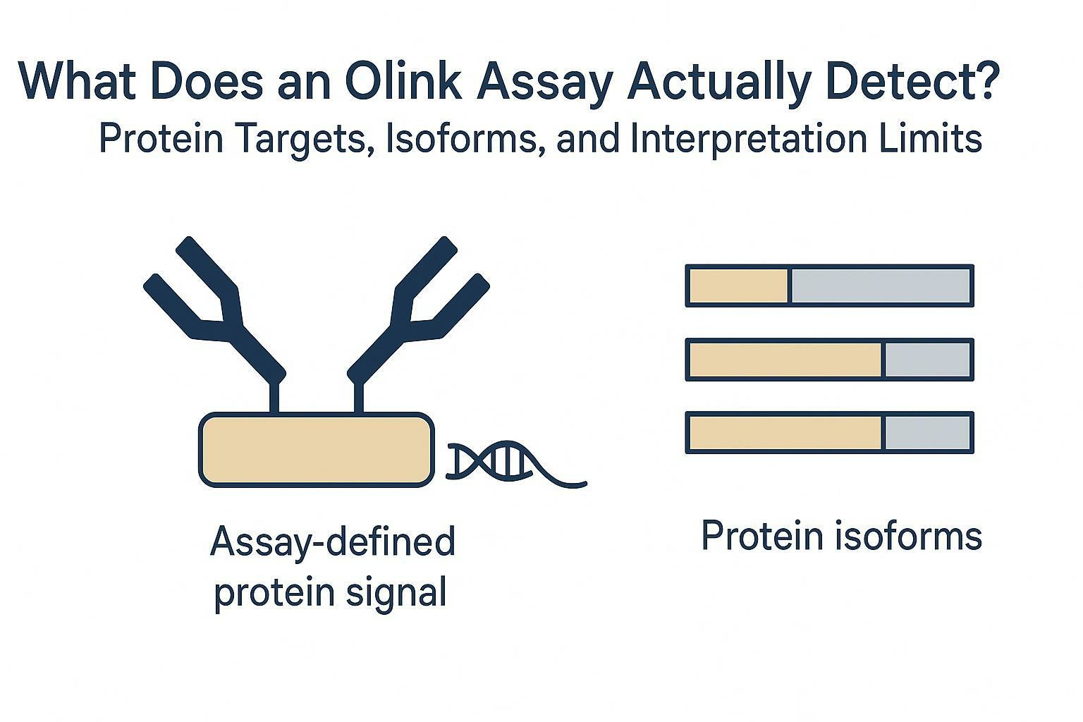
Olink reports an assay-defined protein signal
Olink assays are affinity-based. Signal generation depends on binding and the downstream readout chemistry—not direct protein sequencing. A manuscript-safe framing is:
An Olink readout is best interpreted as an assay-defined protein target signal.
If you need a peer-reviewed description of the proximity-extension concept behind PEA-style assays, see Lundberg et al., Nucleic Acids Research (2011).
NPX is a relative expression readout
In most projects, results are reported as NPX (Normalized Protein eXpression). NPX is commonly treated as a relative, log2-scaled expression value used for within-study comparisons for a given target.
A practical way to state this in study documentation:
- NPX supports relative comparisons across samples for the same assay target.
- NPX is not automatically an absolute concentration (e.g., pg/mL).
- Cross-project comparability requires deliberate design (controls/bridging) and consistent processing.
For wording aligned with Creative Proteomics' workflow (including QC, normalization, LOD handling), see Creative Proteomics' Olink data analysis process.
Protein target ≠ isoform target (by default)
A gene symbol identifies a locus; it does not identify which isoform contributes to the measured signal.
One gene can produce multiple protein isoforms via alternative splicing and related mechanisms. Isoforms can differ in domain composition, termini, and functional regions—exactly the features an affinity assay may (or may not) be sensitive to. For a review overview, see Stamm et al., Gene (2005).
Why Protein Isoforms Complicate Olink Data Interpretation
Isoforms share regions—and assays bind regions
Many isoforms share most of their sequence and differ only by:
- an alternative exon (domain gained/lost)
- a truncated N- or C-terminus
- a different intracellular tail
- a cleavage product vs full-length form
If an assay targets a shared region, the readout may reflect more than one isoform.
Shared regions can produce non-isoform-specific signals
When isoforms share the targeted region, the safest interpretation is:
- "the assay target signal increased"
- "protein target-level expression was higher"
- "the readout may reflect one or more isoforms sharing the targeted region"
Avoid:
- "isoform 1 increased"
- "canonical isoform is elevated"
…unless you can support the target region and specificity.
Why specificity cannot be assumed in multiplex immunoassays
Even with a correct target label, antibody-based multiplex platforms face known constraints: cross-reactivity, nonspecific binding, and interference effects can occur and must be validated in context.
A peer-reviewed reference that's useful for "why validation matters" language:
Related background on immunoassay interference: Tate & Ward, Clinical Biochemistry Review (2004)
Can Olink Distinguish Specific Protein Isoforms?
Olink isoform detection depends on the assay target region
This is the core reason "Olink protein target" labels don't automatically translate into isoform-level statements: the measurement is constrained by what region the assay effectively recognizes.
If you're trying to interpret an Olink signal at isoform resolution, the decision point is whether the assay binds a shared region or a unique region.
Table 1: Shared region vs isoform-specific region interpretation
| Assay target situation | How to interpret the signal | What to do next |
| Shared region across multiple isoforms | Protein target-level signal | Avoid isoform-specific claims; use cautious language |
| Unique isoform region | Isoform-specific interpretation may be possible | Confirm target region + specificity evidence; consider orthogonal validation |
| Target region unclear | Interpretation uncertain | Request technical review; default to target-level interpretation |
| High similarity protein family | Potential specificity concern | Check cross-reactivity evidence; consider orthogonal confirmation |
| Isoform distinction central to hypothesis | Higher confidence required | Plan validation designed to resolve isoforms |
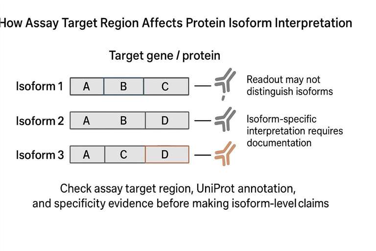 Isoform interpretation depends on whether the assay targets a protein region shared by multiple isoforms or a region unique to one isoform.
Scenario 1: the assay targets a shared protein region
If the assay recognizes a shared region, treat the output as Olink protein expression data for the assay-defined target, not for a specific isoform.
You can still do good science with this signal—just keep the claim aligned to what the assay can support.
Scenario 2: the assay targets a unique region
If the target region is unique to one isoform, isoform-level interpretation becomes more plausible.
However, the claim still hinges on:
- documentation that the assay targets that unique region
- specificity evidence (including how cross-reactivity was evaluated)
- whether your sample matrix and expected abundance range are compatible
Scenario 3: the target region is not clear
If the binding region is not clear, do not guess.
Default interpretation: assay-defined protein signal. If isoforms are central, you should plan orthogonal work.
What Researchers Should Check Before Making Isoform-Specific Claims
This section doubles as an Olink assay interpretation workflow.
1) Confirm target identity (gene symbol + protein name)
This is the foundation for downstream checks. It also reduces confusion when synonyms or legacy names exist.
2) Check UniProt isoforms and annotations
Use UniProt to identify:
- canonical vs alternative isoforms
- shared vs unique regions/domains
- potential cleavage products or processed forms (when relevant)
3) Ask for assay target region / target coverage details
This is the practical heart of Olink assay target coverage questions.
If the service documentation does not disclose the target region, request a technical review. If your decision depends on isoform resolution, make that explicit.
4) Review specificity evidence
When available, look for:
- cross-reactivity testing against related proteins
- validation in a relevant matrix
- clarity on what was and wasn't tested
Multiplex configuration can add complexity; antibodies validated in one context may behave differently in another (Ellington et al., 2010).
5) Decide whether isoform distinction is biologically necessary
A quick decision rule:
- If your conclusion is "this target tracks with phenotype," target-level may be sufficient.
- If your conclusion is "isoform X drives mechanism," you likely need isoform-resolving evidence.
Table 2: What to check before making isoform claims
| Information to check | Why it matters |
| Gene symbol | Confirms target identity |
| Protein name | Aligns assay label with interpretation |
| UniProt entry | Canonical + isoform annotations |
| Target region | Shared vs unique region detection |
| Olink antibody specificity evidence | Supports interpretation; flags cross-reactivity |
| Related proteins | Identifies homolog risks |
| Orthogonal evidence | Strengthens high-stakes claims |
Key Takeaway: If you can't connect the assay to a defensible target region, write results as "assay target signal" and avoid isoform language.
How to Interpret NPX When Isoforms Are Involved
Treat NPX as an assay-specific signal
In isoform-uncertain situations, NPX is best treated as an assay-defined readout for a protein target signal. Keep the interpretation scoped to:
- direction of change
- relative differences within your study design
- QC-aware conclusions
Avoid turning NPX into isoform claims
A common failure mode is converting a protein target signal into a mechanistic isoform claim. If the isoform matters:
- state what the assay can support
- state what would be required to claim isoform specificity
- design orthogonal validation accordingly
When Orthogonal Validation May Be Needed
When isoform-level biology changes the conclusion
If isoforms have different functions, localization, or interaction partners, then "which isoform?" becomes a decision-critical question.
Targeted MS for peptide-level evidence
Targeted MS (SRM/PRM) can measure peptides, and isoform inference may be possible when unique peptides exist and are measurable in your workflow.
For an accessible methods review, see Picotti & Aebersold, Nature Methods (2012).
Western blot / ELISA (when isoform-resolving reagents exist)
Western blot and ELISA are useful when:
- isoforms can be separated (size/processing)
- antibodies are validated for the specific isoform question
Immunoassay interference and cross-reactivity are well-described issues, so validation details matter (see Tate & Ward, 2004).
Practical guidance: match validation to the question
- "Which isoform?" → targeted MS or isoform-specific antibodies (if validated)
- "Is this target real at protein level?" → orthogonal immunoassay or MS, depending on constraints
- "Is the effect robust?" → replicate, confirm QC, and verify the interpretation plan
Technical review before you lock the panel
If isoform interpretation or specificity is decision-critical, it's often worth doing a short technical review before you finalize a panel and sample plan.
Start with:
- Creative Proteomics Olink proteomics assay services
- How to choose the right Olink panel for your research
Practical Example: Handling an MZB1 Isoform-Specific Inquiry (Generic)
A researcher asks: "Does the assay detect the canonical MZB1 isoform, or a region shared across isoforms?"
A careful, defensible approach:
- Confirm target identity (gene symbol/protein name/UniProt entry).
- Check isoform structure (shared vs unique regions).
- Request target region / specificity evidence if not already disclosed.
- State what can be supported (target-level vs isoform-aware language).
- Recommend orthogonal confirmation if the isoform decision changes the interpretation.
Quote / Technical Review Checklist (Pre-order)
Use this checklist to reduce ambiguity before ordering.
- Which gene symbol/protein name and UniProt entry are used for the assay annotation?
- Is the assay target region documented (even at domain/segment level)?
- Does that region overlap a shared domain across isoforms, or a unique segment?
- What evidence supports Olink antibody specificity and cross-reactivity assessment?
- Are there closely related family members with similar domains that could confound interpretation?
- For my sample matrix, what QC and normalization outputs will be provided?
- If I need isoform-level evidence, what orthogonal method do you recommend (targeted MS / isoform-resolving immunoassay), and what sample volume will it require?
Common Mistakes
- Assuming gene symbol equals isoform specificity. Labels aren't regions.
- Treating NPX as absolute concentration. Keep NPX as relative unless you have explicit documentation.
- Overstating specificity. If isoform resolution matters, require documentation and/or orthogonal evidence.
Conclusion
Olink assays can be highly useful for biomarker work—especially when you interpret the output as an assay-defined protein target signal and keep claims aligned to what specificity evidence supports.
If isoform resolution changes the biology, treat it as a study design requirement and build an orthogonal validation plan from the start.
Next step: Share your target list (gene symbols), isoform question, sample type, and study goal. We can help review panel fit and interpretation guardrails before you finalize an Olink panel.
FAQ
Can Olink detect specific protein isoforms?
Sometimes. Olink isoform detection depends on whether the assay targets a region that is unique to the isoform of interest. If the target region is shared across isoforms—or is not clearly documented—interpret the readout at the protein target level and consider orthogonal confirmation for isoform-specific conclusions.
What does NPX represent?
NPX is commonly used as a relative protein expression readout for a given assay target (often log2-scaled). It supports within-study comparisons but should not automatically be interpreted as absolute concentration without panel/workflow documentation.
How do I evaluate biomarker assay specificity in affinity proteomics?
Start with the target region and cross-reactivity evidence. If documentation is insufficient, default to assay-defined interpretation and validate critical claims with an orthogonal method.
About the Author
CAIMEI LI
Senior Scientist at Creative Proteomics
LinkedIn: Caimei Li
Research Use Only (RUO): The content and services described are for research use only and are not intended for diagnostic or clinical decision-making.


