
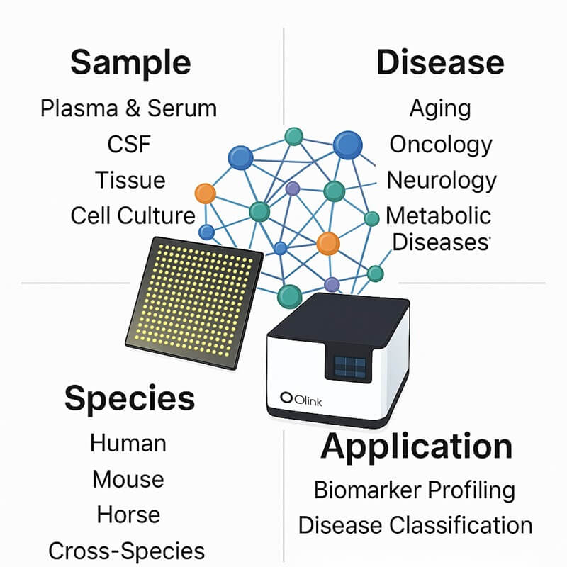
- Panel Features
- Panels List
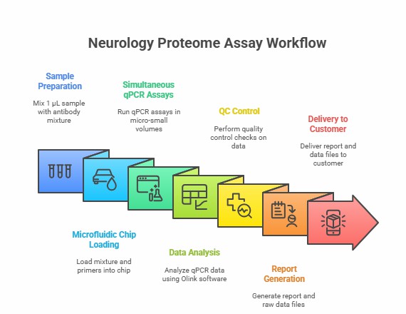
- Workflow
- Why Creative Proteomics
- Demo
- Sample Requirements
- Case
- FAQ
- Sample Submission Pack
Customized and Ready-to-Use Olink Proteomics Panel for Neurology
Why Focus on Neurology Proteomics?
1. Non-Invasive Biomarker Access: Study neurological processes through easily accessible biofluids, enabling longitudinal monitoring without invasive tissue collection
2. Real-Time Pathway Insights: Track dynamic changes in neuroimmune crosstalk, barrier integrity, and glial signaling across experimental conditions
3. Comprehensive Protein Profiling: Capture circulating neurotrophic factors, synaptic proteins, and inflammatory mediators missed in tissue-specific analyses
By leveraging neurology-focused proteomics, researchers gain critical insights into the mechanisms of nervous system communication, thereby accelerating the discovery of biomarkers for neural function, plasticity, and adaptive responses in both physiological and pathological states.
What We Provide
- Disease-focused Olink projects: AD/PD/ALS, MS, stroke/TBI, psychiatric & neurodevelopment, BBB dysfunction, neuropathies.
- Multi-species support: Human (CSF/plasma/serum), mouse/rat; cross-species biomarker mapping.
- Olink + multi-omics: Integrate RNA-seq (bulk/single-cell/spatial), genomics, metabolomics; pathway/network outputs.
- End-to-end setup: 21–384 plex panel selection, SOPs & shipping, longitudinal design, and batch bridging.
- Stats & validation: NPX + QC, differential/longitudinal models, enrichment; ELISA/MSD/LC-MS confirmation.
Optimized for Neuroscience Research
- Neuroinflammatory pathway analysis
- Synaptic function and neural communication studies
- Blood-brain barrier integrity assessment
- Neurodegenerative mechanism investigation
Features of the panel
- Species: Optimized for human proteome profiling in neurological studies
- Design: Customizable target selection via bioinformatics interface for neural pathways
- Proteins 21- to 384-plex panels targeting neuroinflammatory markers, synaptic proteins, and blood-brain barrier components
- Sample: 1-6µL plasma/serum or cerebrospinal fluid per analysis
- Readout: Normalized Protein Expression (NPX) values.
- Platform: PEA technology with high sensitivity and reproducibility.
List of Our Neurology Proteome Assay Panel
Protein category
The Neurology Diseases Proteome Assay Panel quantitatively analyzes proteins across six essential functional categories for neurological research: Neuroinflammatory Regulators, Synaptic Signaling Proteins, Blood-Brain Barrier Components, Neurotrophic Factors, Glial Cell Markers, and Neuronal Structural Proteins (see Table: List of Neurology Biomarker Assay).
This panel encompasses fundamental biological processes, including neural transmission, neuroimmune interactions, blood-brain barrier integrity, synaptic plasticity, and cellular stress responses in neural tissue, featuring strategically selected targets implicated in neurological pathway mechanisms. Through systematic literature curation and experimental validation, we incorporate rigorously vetted biomarkers associated with axon guidance, myelination, neurotransmitter regulation, and glial-neuronal communication, providing researchers with a specialized tool for comprehensive proteome investigations in diverse neurological research areas, including neurodegenerative mechanisms, neural development studies, and neuroinflammatory response monitoring. The panel's optimized design enables focused examination of neurology-related protein interaction networks while preserving the analytical depth required for mechanistic discovery in neuroscience.
Table. List of Neurology Biomarker Assays
Protein Functions
Biological process
primarily involved in key biological processes including synaptic transmission regulation, neuroinflammatory modulation, blood-brain barrier maintenance, axonal transport mechanisms, and glial cell communication, enabling comprehensive investigation of neural pathway dynamics and cellular interactions within the nervous system.
Disease area
primarily utilized in research areas involving neurodegenerative mechanisms, neuroinflammatory models, synaptic plasticity studies, blood-brain barrier dysfunction, and neuronal-glia interaction research, providing investigators with essential tools for exploring fundamental biological mechanisms underlying neurological function and dysfunction.
The Application of Neurology Proteome Assay.
The Neurology Proteome Assay enables high-throughput quantification of protein biomarkers across neural pathways, providing researchers with a powerful tool for:
- Discovery of protein networks underlying synaptic dysfunction and neuroinflammatory responses in neurodegenerative models;
- Mechanistic investigation of blood-brain barrier disruption and glial activation pathways in neurological disorder research;
- Identification of molecular signatures associated with axonal damage and myelin degradation in experimental models
- Analysis of neurotrophic signaling alterations and neuronal-glia crosstalk in neural development studies.
Workflow of Olink Proteomics
Why Creativ Proteomics
Neurology-Specific Analytical Frameworks
Preconfigured workflows for neuroinflammation, synaptic function, and blood-brain barrier studies with 384+ validated biomarker ratios.
Sample Flexibility
Optimized protocols for CSF, plasma, and serum to support diverse neurological study designs.
Longitudinal Study Optimization
Stable normalization controls and batch correction algorithms for long-term neurological monitoring.
Pathway-Centric Bioinformatics
Automated tools for neural pathway enrichment analysis (e.g., synaptic signaling, glial activation).
Demo Results of Olink Data
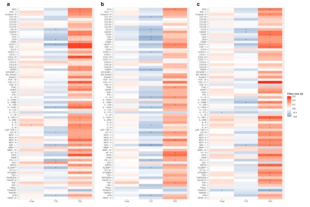 Figure 1: Heatmaps show associations between baseline levels of inflammatory biomarkers and changes in depressive symptom clusters. (Herder, C., et al. 2025)
Sample Requirements
| Sample Type | Recommended Sample Size | Sample Quality | Pre-treatment and Storage | Sample Transport |
| Plasma/Serum/Body Fluid | The minimum input volume per assay is 40 µL. | For optimal results, maintain concentrations between 0.5 and 1 mg/mL. | For long-term preservation, aliquot samples and maintain at -80°C. | Ship sealed samples on dry ice (-80°C). |
| Tissue | ||||
| Cells | ||||
| Exosomes | ||||
| Other |
Case Study
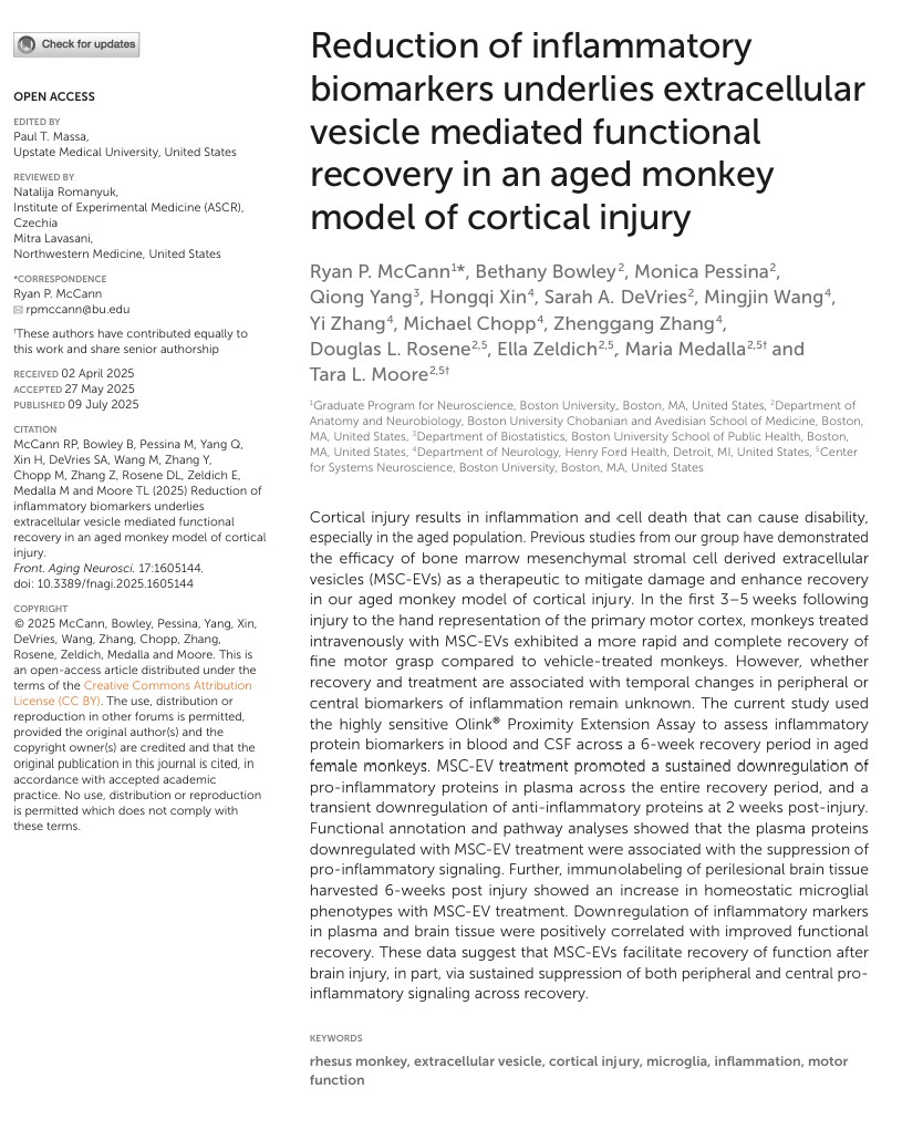
Reduction of inflammatory biomarkers underlies extracellular vesicle mediated functional recovery in an aged monkey model of cortical injury
Journal: Frontiers in aging neuroscience
Year: 2025
- Background
- Methods
- Results
Cortical damage can lead to inflammation and cell death, which can lead to disability, especially in the elderly population. Previous studies have shown that bone marrow mesenchymal stromal cell-derived extracellular vesicles (MSC-EVs) are capable of mitigating damage and promoting recovery as a therapy for our model of cortical damage in aged monkeys. During the first 3 to 5 weeks after injury to the representative area of the motor cortex of the hand, monkeys receiving intravenous injections of MSC-EVs recovered faster and more completely than monkeys receiving control injections. However, it remains unclear whether recovery and treatment are associated with temporal changes in peripheral or central inflammatory biomarkers.
To detect inflammatory protein levels in plasma and cerebrospinal fluid, the collected convalescent samples were tested using the Olink Targeted 96 Inflammation panel. Samples collected from baseline, 24 hours, 2 weeks, 4 weeks, and 6 weeks were sent to the Olink Analysis Center in Waltham, Massachusetts. This multiplex protein measurement assay containing 92 inflammatory proteins uses two antibodies against each protein of interest. Each antibody carries a single-stranded DNA fragment. When two pairs of paired antibodies correctly bind to the protein of interest, the DNA fragments hybridize. The hybridized DNA fragment is stretched to produce a barcoded DNA fragment specific to the protein of interest. The number of encoded fragments in a DNA library is related to the relative concentration of proteins. Positive control samples are used to determine the effect of the test, and negative control samples are used to determine the sensitivity of the test.
To understand the biological effects of injury and MSC-EV treatment during recovery, the investigators collected blood samples at five time points: baseline, 24 hours, 2 weeks, 4 weeks, and 6 weeks during recovery. Quantitative analysis of levels of inflammatory proteins, cytokines, chemokines, neurotrophic factors, and growth factors in plasma was performed using Olink PEA technology. The investigators evaluated the role of injury in the MSC-EV treatment and control groups, comparing the original NPX expression values of each biomarker in each group and time point (Fig.1).
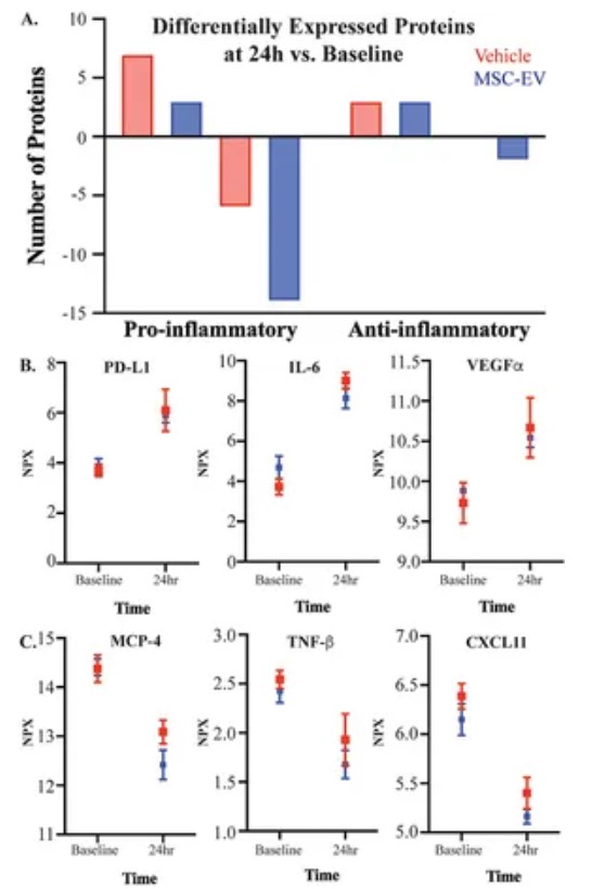 Figure 1. Injury impact on inflammatory plasma profiles. (McCann, R. P. et al. 2025)
FAQs
What sample types are compatible with neurology panels?
The panels support cerebrospinal fluid (CSF), plasma, and serum samples, with optimized protocols for each matrix to ensure reproducibility in neurological studies.
Can panels be customized for specific neurological pathways?
Researchers can select from pre-validated panels or create custom configurations focusing on neuroinflammation, synaptic function, blood-brain barrier integrity, or neurodegeneration pathways.
How are data analyzed for neurological research?
Bioinformatics pipelines provide normalized expression values, pathway enrichment analysis for neural processes, and integration tools for multi-omics correlation studies.
References
- Herder, C., Zhu, A., Schmitt, A., et al. (2025). Biomarkers of inflammation and improvement in depressive symptoms in type 1 and type 2 diabetes: differential associations with depressive symptom clusters. Diabetologia, 68(9), 2057–2068. https://doi.org/10.1007/s00125-025-06472-w
- McCann, R. P., Bowley, B., Pessina, M., et al. (2025). Reduction of inflammatory biomarkers underlies extracellular vesicle mediated functional recovery in an aged monkey model of cortical injury. Frontiers in aging neuroscience, 17, 1605144. https://doi.org/10.3389/fnagi.2025.1605144